Geese Shed Lighting: Effects of Light on the Growth Performance

Directory:
1. Materials and Methods
2. Results and Discussion
3. Discussion
Over the past two decades, the goose farming sector has experienced significant growth, averaging around 4% annually. The prices of goose products have consistently increased, and both the size of breeding populations and the areas dedicated to breeding have expanded, indicating a thriving industry. Recently, the meat goose sector has seen rapid advancements, with large-scale breeding becoming more common. However, a major challenge for breeding companies is how to enhance the production and operational growth of meat geese to achieve higher market weights, which directly impacts their economic returns. Traditionally, geese are raised in aquatic environments, but this method can lead to water pollution and increase the risk of E. coli infections and outbreaks during the summer months. Stricter environmental protection standards necessitate pollution-free, waterless farming, yet geese naturally prefer water. This shift to water-free farming can induce stress and lower disease resistance in geese, leading to decreased production performance and failure to meet market readiness, as well as lower egg production rates. Various factors influence the production performance of geese, such as stocking density, environmental temperature and humidity, air quality, and lighting conditions. Previous research has focused on density issues, and advancements in science and technology have addressed environmental control challenges, with breeders becoming more aware of these factors. Numerous studies have shown that poultry are highly sensitive to light, prompting researchers to investigate how lighting conditions affect poultry production performance.
1. Materials and Methods
1.1 Experimental Materials
1.1.1 Experimental Design and Animals
For this study, 720 healthy 1-day-old geese, averaging around 90 g in weight, were selected from the same batch, consisting of 360 males and 360 females. A 2×2 two-factor crossover design was employed. The meat goose rearing process was categorized into three phases: brooding (1-28 days), rearing (29-50 days), and fattening (51-75 days). The goslings were randomly assigned to four groups, each exposed to different lighting conditions. The lighting treatments included: white light (16 hours light: 8 hours dark) for the control group, white light (22 hours light: 2 hours dark) for one experimental group, green light (16 hours light: 8 hours dark) for another experimental group, and green light (22 hours light: 2 hours dark) for the final experimental group. Each group contained 180 geese, with 90 males and 90 females, and the lighting was maintained throughout the brooding phase. After this period, the groups were expanded, and the remaining goslings were randomly divided into six replicates, ensuring equal male and female representation in each. LED light strips emitting white and green light (wavelength 560 nm) were used as the light source, with an irradiance of 0.1 W/m².
1.1.2 Feeding Management
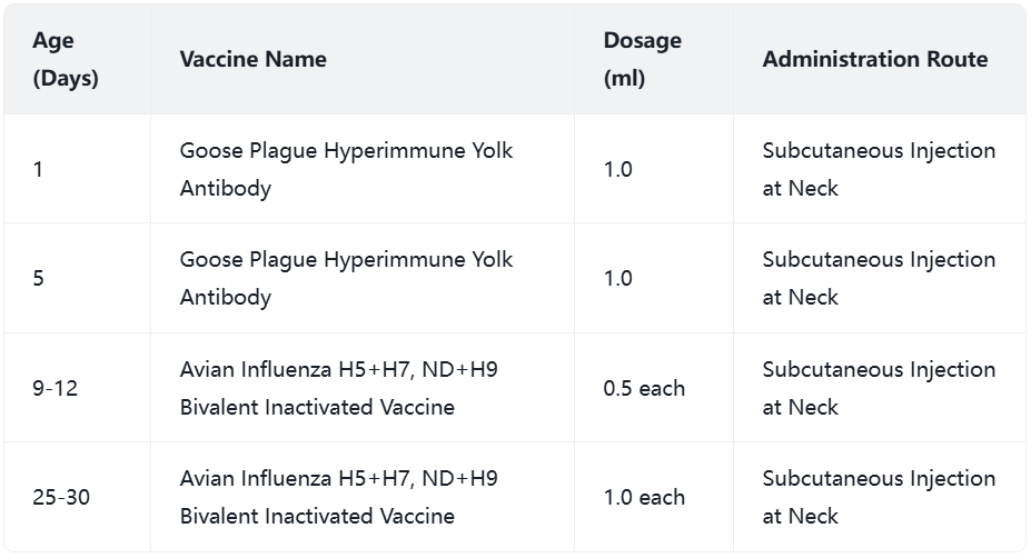
The experiment was conducted at a designated scientific poultry farm, with the geese sourced from a specific goose industry company. Two identical goose houses were utilized, each measuring 36 m in length and 12 m in width, separated into two compartments by black shading cloth. Each compartment was further divided into six equally sized pens. The geese were raised on elevated frames made of slatted plastic boards, positioned 0.75 m above the ground. Temperature and humidity were maintained consistently across all groups, controlled automatically by heaters and fans. During the experiment, the houses were kept closed to prevent natural light from entering, and the geese had unrestricted access to food and water. The effects of the monochromatic light treatments are illustrated in Figure 3. while the immunization schedule is detailed in Table 1. The total duration of the experiment was 75 days.
Figure 3 LED green and white light processing renderings

Table 1 Immunization procedures

1.1.3 Key Instruments and Reagents
Key instruments include: LED light strip, heater, weighing scale, electronic scale, surgical tools, blood collection tubes, liquid nitrogen, collection tubes, culture dishes, and physiological saline.
Key reagents consist of: potassium permanganate, formaldehyde, serum nutrition index kit, and serum biochemical index ELISA kit.
1.2 Testing Methods
1.2.1 Testing Parameters and Sample Collection
At 10. 20. 30. 50. and 75 days of age, one goose (6 from each group, with 3 males and 3 females) was randomly chosen from each group and replicate for weighing and blood collection from the wing vein. The goose was then euthanized by bleeding from the posterior carotid artery after administering urethane (1g/kg). The geese were not fed for 12 hours prior to slaughter. After dissection, the heart, spleen, liver, left pectoral muscle, and left leg muscle were collected and weighed. Additionally, 3 grams each of the hypothalamus, pituitary gland, liver, and pectoral muscle were placed in cryopreservation tubes and immediately stored in liquid nitrogen. For future molecular tests, blood was collected in an anticoagulated vacuum tube and centrifuged at 3000 rpm for 15 minutes at room temperature. The supernatant was carefully extracted and stored in aliquots at -20 ℃.
1.2.2 Detection of Growth Performance Index and Method
Throughout the experiment, the daily feed intake of the geese was monitored, and their fasting weights were measured on the 1st, 10th, 20th, 30th, 40th, 50th, 60th, and 75th days. This data was used to calculate the average daily feed intake, average live weight, and feed-to-weight ratio for each group. The calculations are as follows:
Average live weight: average live weight = total weight of geese in each treatment group / number of geese
Average daily feed intake (ADFI): average daily feed intake = total feed consumed by each treatment group during the experimental period / number of days in the experimental period
Feed-to-weight ratio (F/G): feed-to-weight ratio = feed consumption / body weight gain
1.2.3 Detection of Slaughter Performance Index and Method
Live weight is defined as the weight after a 12-hour fasting period prior to slaughter.
Chest muscle rate = (left chest muscle weight * 2 / total eviscerated weight) * 100%
Leg muscle rate = (left leg net muscle weight * 2 / total eviscerated weight) * 100%
1.2.4 Detection of Organ Index and Method
Organ index (g/kg) = organ weight (g) / live weight before slaughter (kg)
1.2.5 Detection of Blood Biochemical Indices and Methodology
Following the guidelines of the standard test kit, serum levels of TG, T-CHO, and GLU were measured using an enzyme marker. For GH, IGF-1. T3. and T4 levels, a fully automated biochemical analyzer was utilized in accordance with the ELISA test kit.
1.3 Data Analysis and Statistics
Experimental data were processed using Excel 2013. while SPSS 19.0 was employed for differential analysis. One-way ANOVA, along with LSD and Duncan’s multiple comparison two-way ANOVA, were conducted, and results were presented as mean ± standard deviation (Mean+SD).
2. Results and Discussion
2.1 Effect of Various Light Conditions on the Growth Performance of Geese
The weight of meat geese was influenced by different light conditions. As illustrated in Figure 4 and Table 2. at 40 days old, the body weight of the group exposed to green light for 22 hours was significantly greater than that of the group exposed to white light for 16 hours (P<0.05). No significant differences were observed among the other two groups. The green light treatment group consistently showed higher body weight compared to the white light group, with significant differences noted (P<0.05). The 22-hour treatment group also had a significantly higher body weight than the 16-hour group (P<0.05). The interaction between light color and duration did not significantly affect the outcomes. At 50 days, a significant difference in body weight was noted between the 22-hour and 16-hour treatment groups (P<0.05). By 60 days, the green light 22-hour treatment group again showed a higher body weight than the white light 16-hour group, with significant differences (P<0.05). The green light treatment group maintained a higher body weight compared to the white light group, with significant differences (P<0.05). At 75 days, significant differences in body weight were observed among the treatments. The 22-hour treatment group did not differ significantly from the 22-hour white light group, but did differ significantly from the two green light treatment groups (P<0.05). The 16-hour green light treatment group did not show a significant difference from the 22-hour green light treatment group. Two-factor analysis indicated that the body weight gain of the green light treatment group was significantly different from that of the white light treatment group (P<0.01), and the body weight of the 22-hour treatment group was significantly different from that of the 16-hour treatment group (P<0.05). The interaction between light color and duration did not significantly impact the various treatment groups.
Figure 4 Effects of different light factors on average live body weight of geese

According to Table 3. there was no significant difference in the feed intake of meat geese based on varying light conditions (P>0.05). Throughout all phases, the group exposed to 22 hours of light consumed more feed than the group with 16 hours of light, although this difference was not statistically significant. Additionally, neither the color nor the duration of light, nor their interaction, had a significant impact on the different treatments.
Table 3 Effects of different light factors on feed intake of geese

Various lighting conditions impact the feed-to-weight ratio of meat geese differently. According to Table 4. from 31 to 50 days old, there was no notable difference in the feed-to-weight ratio among the treatments. However, from 51 to 75 days old, the group exposed to white light had a significantly higher feed-to-weight ratio compared to the group exposed to green light (P<0.05). The duration of light and its interaction did not significantly affect the feed-to-weight ratio across the treatments. Over the entire period from 31 to 75 days, there were no significant differences in the feed-to-weight ratios among the treatments. Nonetheless, it was observed that the group receiving 22 hours of green light had the lowest feed-to-weight ratio, followed by the group with 16 hours of green light, then the group with 16 hours of white light, while the group with 22 hours of white light had the highest feed-to-weight ratio of 2.81±0.27.
Table 4 Effects of different light factors on feed intake of geese

2.2 Effect of Various Light Conditions on the Slaughter Performance of Geese
The slaughter performance of geese is significantly affected by different light conditions. As indicated in Table 5. the group exposed to green light for 22 hours had the highest live weight, which was notably different from the group exposed to white light (P<0.01). When compared to the group with 16 hours of green light, the 22-hour group also showed a significantly higher weight (P<0.01). The weight difference between the green light group and the white light group was highly significant (P<0.01), as was the difference between the 22-hour and 16-hour light treatment groups (P<0.01). Although there were no significant differences in chest muscle weight among the groups, the 16-hour green light group had the heaviest chest muscles, followed by the 22-hour green light group. The chest muscle rate was highest in the 22-hour green light group, but the differences among the groups were not significant. The 16-hour green light group had the heaviest leg muscles, while the highest leg muscle rate was observed in the 16-hour white light group.
Table 5 Effects of Different Light Factors on Slaughter Performance of Geese

2.3 Effect of Various Light Factors on the Organ Index of Geese
Table 6 illustrates that different light factors influence the liver index of geese in distinct ways. At 20 days old, there are significant differences in the liver index among the treatment groups. The group exposed to 16 hours of white light has the highest liver index, surpassing the group exposed to 16 hours of green light (P<0.01) and the other two groups (P<0.05). There is a notable difference between the white light and green light treatment groups (P<0.05). The interaction between light color and duration significantly affects the treatments (P<0.05). At 30 days, this interaction continues to have significant effects on the groups (P<0.05). By 50 days, the green light treatment group shows a significant difference from the white light treatment group (P<0.05). At 75 days, the influence of white light on the liver index is significantly different from that of green light (P<0.05).
Table 6 Effects of different light factors on liver index of geese

Various light conditions impact the spleen index of geese differently, as indicated in Table 7. At 20 days of age, there was a significant difference in the spleen index between the green light treatment group and the white light treatment group (P<0.05).
Table 7 Effect of different light factors on spleen index of geese

Various light conditions have distinct impacts on the heart index of geese, as indicated in Table 8. At 20 days of age, the group exposed to 16 hours of white light showed a significant difference compared to the group exposed to 16 hours of green light (P<0.01), as well as compared to other treatment groups (P<0.05). By 50 days of age, the spleen index in the green light treatment group was significantly higher than that in the white light treatment group (P<0.05). Additionally, the interaction between the color of light and its duration significantly influenced the spleen index across the treatments (P<0.05).
Table 8 Influence of different light factors on heart index of geese

2.4 Effect of Various Light Factors on Serum Hormone Levels in Geese
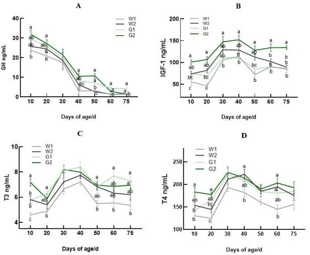
Figure 5 illustrates that the serum growth hormone (GH) levels across the four groups exhibited a consistent decline as the geese aged. The lowest serum GH level was recorded on the 75th day of the experimental period, while the highest was observed on the 10th day. Significant differences among the groups were noted at all ages except for 30 days (P<0.05). At 10 days, group G2 had a notably higher GH level compared to group W1 (P<0.05). By 20 days, group G2's GH level surpassed those of groups W1 and W2. showing significant differences (P<0.05). No significant differences were found among the groups at 30 days (P>0.05). At 40 days, group G2's GH level was significantly higher than that of group W2 (P<0.05) and extremely higher than group W1 (P<0.01). The GH levels at 50 days mirrored those at 40 days. At 60 and 75 days, group W1's GH levels were lower than those of the other three groups (P<0.05). The figure also indicates that serum IGF-1. T3. and T4 levels in all groups initially rose and then fell with age. The serum IGF-1 levels ranked from highest to lowest were G2. W2. G1. and W1. The trends for T3 and T4 levels were largely similar.
Figure 5 Effects of light on serum hormone levels of geese

2.5 Effect of Various Light Factors on Serum Biochemical Parameters of Geese
Table 9 illustrates the influence of different light factors on the routine serum biochemical parameters of geese. At 20 days old, the glucose levels in the green light 22-hour treatment group were significantly higher than those in the 16-hour treatment group (P<0.05), and also higher than in the white light treatment group (P<0.05). Additionally, the 22-hour treatment group had significantly higher glucose levels compared to the 16-hour group (P<0.05). By 30 days of age, the glucose content in the green light 22-hour group surpassed that of the white light 22-hour group (P<0.05), while the 16-hour light treatment group had higher levels than the 22-hour group (P<0.05). At 40 days, the white light 16-hour treatment group exhibited lower glucose levels than the other three groups (P<0.05), with significant differences noted between the green and white light groups (P<0.05), and the 22-hour treatment group showing significantly higher levels than the 16-hour group. The 5-hour treatment group also had significantly different levels compared to the white light group, and there were significant interactions between light color and treatment duration across the groups (P<0.05). At 50 days, significant interactions between light color and treatment duration were observed (P<0.05). By 60 days, the glucose levels in the white light 75-hour treatment group were higher than in the other three groups (P<0.05), with an extremely significant effect of the white light treatment on the green light group (P<0.01). The 16-hour treatment group had higher glucose levels than the 22-hour group (P<0.05), and there were extremely significant interactions between light color and treatment duration (P<0.01).
Table 9 Effects of monochromatic light on serum glucose content of geese

Table 10 illustrates the impact of various light conditions on serum triglyceride levels, a standard biochemical measure for geese. In the 10-day-old group exposed to 22 hours of light, triglyceride levels were significantly higher compared to the 16-hour light group (P<0.05). By 60 days of age, the 16-hour green light group showed significantly elevated levels compared to the 22-hour green light group (P<0.05), with no significant differences noted among the other two groups. Additionally, the white light treatment group had higher levels than the green light group, with a significant difference (P<0.05). The combination of light color and duration significantly influenced the treatment differences. At 75 days old, the green light treatment group had significantly higher levels than the white light group (P<0.01), and the 16-hour light group surpassed the 22-hour light group (P<0.01). The interaction between light color and duration had a substantial effect on the treatment differences (P<0.01).
Table 10 Effects of monochromatic light on serum triglyceride content of geese

Table 11 illustrates the impact of various light factors on total cholesterol, a standard biochemical marker in the serum of geese. At 10 and 20 days old, the group exposed to 22 hours of white light had higher cholesterol levels than the group exposed to 22 hours of green light (P<0.05). Additionally, the group with 16 hours of light showed higher levels than the 22-hour group, with significant differences (P<0.05). The interaction between light color and duration had a highly significant effect on the results (P<0.01). By 30 days of age, the 22-hour green light group had significantly lower cholesterol levels compared to the other three groups (P<0.05). The white light group had higher levels than the green light group, with significant differences (P<0.05), and the 16-hour light group was higher than the 22-hour group, showing extremely significant differences (P<0.01). The interaction between light color and duration again had a highly significant impact (P<0.01). At 40 days, the 16-hour white light group differed significantly from both the 22-hour white light and 16-hour green light groups (P<0.05), and showed extremely significant differences from the 22-hour green light group (P<0.01). The white light group was also extremely significantly different from the green light group (P<0.01), and the 16-hour light group was extremely significantly different from the 22-hour light group (P<0.01). At 50 days, the 22-hour green light group had lower cholesterol levels than the other three groups (P<0.05), with the white light group showing significant differences from the green light group (P<0.01) and the 16-hour light group differing significantly from the 22-hour group (P<0.01). The interaction between light color and duration significantly affected the treatment differences (P<0.01). At 60 days, the 22-hour white light group differed significantly from both the 16-hour and 22-hour green light groups, while the 16-hour light group was significantly different from the white light group (P<0.01). The interaction between light color and duration also significantly influenced the treatment differences. Finally, at 75 days, univariate analysis revealed that the white light group had higher cholesterol levels than the green light group, with a significant difference (P<0.05), and the 16-hour light group was significantly higher than the 22-hour group, with extremely significant differences (P<0.01). The interaction between light color and duration had an extremely significant effect on the outcomes of each treatment group (P<0.01).
Table 11 Effects of monochromatic light on serum total cholesterol content of geese

3. Discussion
3.1 Effect of Various Light Factors on the Growth Performance of Geese
Key indicators for assessing poultry production performance include body weight gain, feed intake, and feed conversion rate. Research by Li Yunlei et al. indicated that applying blue light during the early growth phase enhanced average daily weight gain and overall production performance throughout the growth period. Li Hanqing investigated the effects of white, red, green, and blue LED lights on the growth performance of geese. The findings revealed that green light significantly boosted growth during both the growing and fattening phases, while blue light notably enhanced growth during the middle growing phase (30-50 days old). However, the growth-promoting effect of blue light diminished during the fattening stage, although it resulted in the best meat quality among the four light treatments. It is hypothesized that blue light positively influences the meat quality of geese in later stages. Hassan et al. examined the effects of yellow, green, blue, and white LEDs on the growth performance, blood biochemical markers, bone density, and meat fatty acids of Cherry Valley ducks. They discovered that green light increased weight throughout the experimental period, while yellow light promoted weight gain in the 21 days leading up to the experiment. Between days 22 and 42 of the study, the blue and green light groups showed significant weight gain differences compared to other groups. Rozenboim et al. found that broilers raised under blue and green LED lights had significantly higher weights than those under white and red LED lights. Conversely, Kim et al. reported no significant weight differences among broilers exposed to blue, green, red, and white lights. The varying results across different studies suggest that outcomes may differ among species.
The experiment revealed that geese marketed at 75 days of age experienced significantly greater weight gain under green light conditions compared to those under white light. Additionally, the group exposed to 22 hours of light had a higher average weight than the group with 16 hours of light, aligning with findings from Li Hanqing's research. While there was no difference in overall feed intake among the treatment groups, the 22-hour light group consumed more feed than the 16-hour group, suggesting that longer light exposure can enhance feed intake. The study also indicated that the feed-to-meat ratio for the green light group during the fattening phase was significantly lower than that of the white light group, with the 22-hour green light group having the lowest ratio throughout the trial. This suggests that green light can effectively reduce the feed-to-meat ratio, and extending light duration can further optimize it. In conclusion, utilizing green light and increasing light duration can boost geese weight gain and improve feed efficiency, allowing for greater growth potential at market time, meeting market demands, and enhancing economic returns.
3.2 Effect of Various Lighting Factors on the Slaughter Performance of Geese
Slaughter performance serves as a crucial metric for assessing the growth and quality of livestock and poultry, with the actual meat yield from geese being a significant concern for both consumers and producers. Key indicators such as the half-eviscerated rate, breast muscle rate, and leg muscle rate provide insights into the quality of poultry meat. In modern poultry production, lighting strategies have become essential for regulating growth. Research by Halevy et al. examined how different monochromatic light treatments affect muscle growth and satellite cell proliferation in broilers. Their findings indicated that the green and blue light groups exhibited greater muscle weight due to increased satellite cell proliferation shortly after birth. Specifically, under green light, the number of satellite cells per gram of breast muscle tissue was double that of the control group exposed to white light. Liu et al. demonstrated that green light enhances the development of satellite cell populations in poultry breast muscle, thereby facilitating growth. Liu Wenjie and colleagues investigated the effects of white, red, green, and blue LED lights, with a lighting duration of 23 hours, on broiler muscle growth, muscle fiber development, and serum testosterone levels. Their results revealed that at 21 days of age, the breast and leg muscles of the green light group were 6.46%-13.57% and 6.73%-16.34% larger than those of the other groups, showing significant differences (P<0.05); additionally, the muscle fiber area in the green light group was 23.19%-54.01% greater than in the other groups (P<0.05).
The slaughter results at 75 days of age indicated that the breast muscle weight, breast muscle rate, and leg muscle weight in the 16-hour green light treatment group were significantly higher than those in the 16-hour white light treatment group. This suggests that green light may promote the proliferation and differentiation of satellite cells, thereby enhancing growth performance, which aligns with previous research findings. In conclusion, various lighting factors significantly affect the slaughter performance of poultry, with different slaughter trait indicators varying under distinct lighting conditions.
3.3 Effect of Various Light Factors on the Organ Index of Geese
The heart serves as the core of bodily functions, facilitating blood circulation and delivering nutrients throughout the body to sustain life. The liver, being the largest digestive gland, is crucial for the metabolic processes of the body's three main nutrients. The spleen, the largest peripheral lymphoid organ in birds, is vital for both cellular and humoral immunity. Under normal conditions, the relative weight of these organs can indicate the physiological health of poultry; a higher relative weight often suggests better physiological function. In chickens, the primary immune organs include the thymus, bursa of Fabricius, bone marrow, and Harder's gland. The ratio of the mass (in grams) of the thymus, spleen, and bursa of Fabricius to the live weight (in kilograms) is commonly used as a straightforward measure of broiler immunity. In this study, the immune response of meat geese across different treatment groups was assessed by examining the heart, liver, and spleen. The findings indicated that neither the color of light nor the extension of light duration significantly affected the heart index, nor was there a notable difference in the liver and spleen indices. Consequently, exposure to green light and longer light durations does not enhance the immune performance of meat geese.
3.4 Effect of Various Light Conditions on Blood Biochemical Markers in Geese
Growth hormone-releasing hormone (GHRH) is a polypeptide hormone produced by the hypothalamus and is crucial for stimulating the secretion of growth hormone (GH). It aids in animal growth by enhancing the synthesis and release of GH. This research indicates that serum GH levels tend to decrease as geese age, and there is a positive correlation between serum GH levels and growth rates. Exposure to green light significantly elevates serum GH levels compared to white light, thus facilitating growth. Extending the duration of light exposure can further enhance GH secretion, although the effect is minimal. Insulin-like growth factor 1 (IGF-1) contributes to growth by encouraging the proliferation and differentiation of skeletal muscle satellite cells, and its impact on muscle development is influenced by blood T3 levels. Hypothyroidism can lower IGF-1 levels in rat muscle, hindering muscle growth. The serum IGF-1 levels initially rise and then fall as meat geese age, and this is not consistently correlated with growth rates. In this study, serum IGF-1 levels increased and then decreased with age, peaking at 30 and 40 days. The group exposed to 22 hours of green light had significantly higher IGF-1 levels at all ages compared to the other three groups, followed by the 22-hour white light group, then the 16-hour green light group, with the 16-hour white light group showing the lowest IGF-1 levels. Thus, it is concluded that 16 hours of green light significantly boosts serum IGF-1 levels compared to white light, and extending the exposure to 22 hours further increases IGF-1 levels, promoting satellite cell proliferation.
Thyroid hormones, primarily T4 and T3. are produced by the thyroid gland and play a crucial role as endocrine regulators in various metabolic processes in poultry. These hormones are involved in embryonic development, hatching, bone and muscle growth, temperature regulation, neural development, and feather growth. T3 and T4 are significant growth enhancers, positively affecting skin health and influencing feed intake in chickens. They are also important for regulating growth inhibition and promoting compensatory growth in chicks. In this research, both T3 and T4 exhibited similar patterns, with the green light treatment group showing higher levels than the white light group. Green light appears to enhance the synthesis and secretion of T3. thus supporting growth and metabolism.
The study revealed that monochromatic green light can lower serum total cholesterol levels, likely due to its stimulation of the anterior hypothalamus, which regulates the parasympathetic nervous system and influences bile secretion. Since bile is responsible for breaking down most of the body's cholesterol, this leads to a reduction in total cholesterol levels. While green light treatment was found to increase glucose levels, the differences between treatments were not statistically significant.
Additionally, green light treatment was shown to boost the slaughter weight of geese at 75 days old. Extending the lighting duration to 22 hours further enhanced slaughter weight, decreased the feed-to-meat ratio in the later stages of the experiment, increased serum levels of GH, IGF-1. T3. and T4. reduced total cholesterol levels, and promoted the growth of meat geese.